|
||||||||
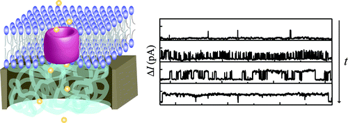
Several nanoporous platforms were functionalized with pH-responsive poly(methacrylic acid) (PMAA) brushes using surface-initiated atom transfer radical polymerization (SI-ATRP). The growth of the PMAA brush and its pH-responsive behavior from the nanoporous platforms were confirmed by scanning electron microscopy (SEM), Fourier transform infrared (FTIR) spectroscopy, and atomic force microscopy (AFM). The swelling behavior of the pH-responsive PMAA brushes grafted only from the nanopore walls was investigated by AFM in aqueous liquid environment with pH values of 4 and 8. AFM images displayed open nanopores at pH 4 and closed ones at pH 8, which rationalizes their use as gating platforms. Ion conductivity across the nanopores was investigated with currentāvoltage measurements at various pH values. Enhanced higher resistance across the nanopores was observed in a neutral polymer brush state (lower pH values) and lower resistance when the brush was charged (higher pH values). By adding a fluorescent dye in an environment of pH 4 or pH 8 at one side of the PMAA-brush functionalized nanopore array chips, diffusion across the nanopores was followed. These experiments displayed faster diffusion rates of the fluorescent molecules at pH 4 (PMAA neutral state, open pores) and slower diffusion at pH 8 (PMAA charged state, closed pores) showing the potential of this technology toward nanoscale valve applications. |
|
||||||||
Surface-bound self-assembled lipid nanotubes (LNTs) made of 1,2-dioleoyl-sn-glycero-3-phosphoethanolamine (DOPE) were used to visualize the contractile activity of spreading cells. The interaction of cells with LNTs resulted in the nucleation of new nanotubes, directed toward the cell center, from existing ones. This process depended on cell generated forces and required acto-myosin mediated contractility. The dynamics of de novo generation of LNTs upon cell spreading was captured using optical microscopy on fluorescently labeled nanotubes and revealed characteristic fingerprints for different cell types such as fibroblasts, endothelial and melanoma cells. Additionally, the method was applied to detect the effect of a specific inhibitor on the generation of cellular forces. The mechanism of the LNTācell interaction and the potential applications are discussed. |
|
||||||||
Conventional lipid-tube formation is based on either a tube phase of certain lipids or the shape transformation of lamellar structures by applying a point load. In the present study, lipid blocks in inverted hexagonal phase made of 1,2-dioleoyl-sn-glycero-3-phosphoethanolamine (DOPE) were shown to protrude lipid nanotubes upon a fluid-dynamic flow on polyelectrolyte-functionalized surfaces in physiological buffer solution. The outer diameter of the tubes is 19.1 Ā± 4.5 nm and their lengths are up to several hundred micrometers. The method described enables the alignment and patterning of lipid nanotubes into various (including curvy) shapes with a microfluidic system. |
|
||||||||
Inverted hexagonal blocks of 1,2-dioleoyl-sn-glycero-3-phosphoethanolamine (DOPE) lipid adsorbed on a polyethyleneimine (PEI)-coated surface in deionized water transformed its shape upon the application of an electric field, forming lipid objects in a variety of shapes (e.g. lines with a width of 10ā50 Ī¼m). The phenomenon was driven by the electrophoresis, because the zwitterionic lipid, DOPE turned out to be highly negatively charged in deionized water. The interaction between DOPE and the PEI surface stabilized the system, assuring a lifetime over several weeks for the formed structures after the electric field was switched off. The free-drawing of microscopic objects (lines, crosses, and jelly fish) was also achieved by controlling the direction of the lipid movement with the field direction. |
|
||||||||
A procedure based on freezing and thawing was developed to induce the rupture of adsorbed lipid vesicles on solid surfaces into supported lipid bilayers (SLBs). The SLB assembly exploits the phase transition of both lipids and water during freezing. It enables SLB formation independent of the type of substrates and lipids as long as the vesicles spontaneously adsorb onto the surface. The created SLB is a single bilayer, and has a diffusion coefficient of (0.6ā4) Ć 10ā8 cm2 sā1 on TiO2, which is in the same range as the SLBs formed by conventional techniques. The presented approach has the advantages of both the LangmuirāBlodgett method (the versatility in the selection of lipids and substrates) and vesicle fusion (self-assembly) at the same time. |
|
||||||||
The enormous progress of nanotechnology during the last decade has made it possible to fabricate a great variety of nanostructures. On the nanoscale, metals exhibit special electrical and optical properties, which can be utilized for novel applications. In particular, plasmonic sensors including both the established technique of surface plasmon resonance and more recent nanoplasmonic sensors, have recently attracted much attention. However, some of the simplest and most successful sensors, such as the glucose biosensor, are based on electrical readout. In this review we describe the implementation of electrochemistry with plasmonic nanostructures for combined electrical and optical signal transduction. We highlight results from different types of metallic nanostructures such as nanoparticles, nanowires, nanoholes or simply films of nanoscale thickness. We briefly give an overview of their optical properties and discuss implementation of electrochemical methods. In particular, we review studies on how electrochemical potentials influence the plasmon resonances in different nanostructures, as this type of fundamental understanding is necessary for successful combination of the methods. Although several combined platforms exist, many are not yet in use as sensors partly because of the complicated effects from electrochemical potentials on plasmon resonances. Yet, there are clearly promising aspects of these sensor combinations and we conclude this review by discussing the advantages of synchronized electrical and optical readout, illustrating the versatility of these technologies. |
|
||||||||
A simultaneous optical waveguide lightmode spectroscopy (OWLS) and electrochemical impedance spectroscopy (EIS) measurement was carried out for the investigation of a supported lipid bilayer and its interactions with a pore-forming peptide, melittin. It was achieved only after the optimization of the ITO coating on the waveguide to increase the electrical sensitivity and the functionalization of the waveguide with a polyelectrolyte to form a lipid bilayer over the ITO surface. The combined system enabled monitoring of melittin pore activities in a wider range of melittin concentrations than either technique alone (1 Ī¼g/ml < Cmelittin < 200 Ī¼g/ml). Furthermore, it provided unique information that could not be obtained by the individual methods, such as a better identification of the melittin-pore formation and an insight about the correlation between the total pore area vs. adsorbed amount of melittin. |
|
||||||||
This review describes and discusses techniques useful for monitoring the activity of protein ion channelsin vitro. In the first section the biological importance and the classification of ion channels are outlined in order to justify the strong motivation for dealing with this important class of membrane proteins. The expression, reconstitution and integration of recombinant proteins into lipid bilayers are crucial steps to obtain consistent data when working with ion channels. In the second section recording techniques used in research are presented. Since this review focuses on analytical systems bearing reconstituted ion channels the industrial most important patch-clamp techniques of cells are only briefly mentioned. In section three, artificial systems developed in the last decades are described while the emerging technologies using nanostructured supports or microfluidic systems are presented in section four. Finally, the remaining challenges of membrane protein analysis and its potential applications are briefly outlined. |
|
||||||||
The resistivity ĻPEM of polyelectrolyte multilayers (PEMs), PEI(PSS/PAH)24, PEI(PGA/PAH)12, PEI(HA/PLL)12 and PEI(PSS/PLL)12, in a free-hanging configuration was estimated combining electrochemical impedance spectroscopy (EIS) and atomic force microscopic (AFM) images. Surprisingly, the obtained value of several kĪ©cm is at least 6 orders of magnitude lower than that reported previously, where the resistivity was determined in the conventional PEM-on-electrode system. The significant discrepancy indicates the unexpectedly low electrical PEM resistance in the absence of redox-active ions and the sensitivity limitation in the conventional system. |
|
||||||||
Sensitive and selective biosensors for high-throughput screening are having an increasing impact in modern medical care. The establishment of robust protein biosensing platforms however remains challenging, especially when membrane proteins are involved. Although this type of proteins is of enormous relevance since they are considered in >60% of the pharmaceutical drug targets, their fragile nature (i.e., the requirement to preserve their natural lipid environment to avoid denaturation and loss of function) puts strong additional prerequisites onto a successful biochip. In this review, the leading approaches to create lipid membrane-based arrays towards the creation of membrane protein biosensing platforms are described. Liposomes assembled in micro- and nanoarrays and the successful set-ups containing functional membrane proteins, as well as the use of liposomes in networks, are discussed in the first part. Then, the complementary approaches to create cell-mimicking supported membrane patches on a substrate in an array format will be addressed. Finally, the progress in assembling free-standing (functional) lipid bilayers over nanopore arrays for ion channel sensing will be reported. This review illustrates the rapid pace by which advances are being made towards the creation of a heterogeneous biochip for the high-throughput screening of membrane proteins for diagnostics, drug screening, or drug discovery purposes. |
|
||||||||
A lipid bilayer with gigaohm resistance was fabricated over a single 800 nm pore in a Si3N4 chip using 50 nm liposomes. The nanopore was prefilled with a polyelectrolyte multilayer (PEM) that triggered the spontaneous fusion of the lipid vesicles. Pore-forming peptide melittin was incorporated in the bilayer, and single channel activities were monitored for a period of 2.5 weeks. The long lifetime of the system enabled the observation of the time-dependent stabilization effect of the melittin open state upon bias application. |